특발성호흡곤란 증후군(특발성 호흡장애 증후군/유리질막증) Idiopathic respiratory distress syndrome in newborn infants(IRDS)

posted in: Uncategorized | 0

특발성호흡곤란 증후군(특발성 호흡장애 증후군/유리질막증), Idiopathic respiratory distress syndrome in newborn infants(IRDS)

다음 정보를 참조 www.drleepediatrics.com-제6권 성장 발육 양육 질병   26장 : 신생아의 호흡기의 질환

Please visit www.drleepediatrics.com-Volume 6  growth development parenting diseases -chapter 26: newborn respiratory Disease
201.jpg

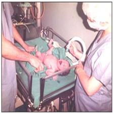
사진 307.미숙아로 태어난 신생아가 호흡곤란이 생겨 산소 튜브로  산소호흡 치료받고 있다.

그 후 특발성 호흡곤란 증후군을 진단받고 치료를 받았다.

Copyright ⓒ 2011 John Sangwon Lee, M.D., FAAP

  • 아주 작은 미숙아들의 폐는 더 미숙해서 만삭에 태어난 건강한 신생아들의 폐에 비해서 생리적‧기능적으로 특이한 점이 많다. 수정체 후 섬유증식증 참조.
  • 미숙아 폐기포(肺氣胞/폐포) 내에 서팩탄트(표면활성제/Surfactant)라는 생화학적 물질이 정상적으로 충분히 들어 있지 않다.
  •  폐포와 세기관지(細氣管支/모세기관지)도 더 미숙하고 그 기능도 미숙하다.
  • 흡입된 공기 속 산소와 몸속에 생긴 이산화탄소가 미숙아의 폐포에서 적절히 잘 교환되지 않는다.
  • 이런 여러 가지 이유로 특히 아주 작은 미숙 신생아의 폐에 특발성 호흡곤란 증후군 등 여러 가지 병이 더 잘 생길 수 있다.
  • 폐가 미숙하기 때문에 생기는 병들 중 유리질막증(특발성 호흡곤란 증후군)이 미숙 신생아에게 더 잘 생길 수 있다.
  • 과거에는 특발성 호흡곤란 증후군은 미숙아들의 사망의 주원이다.
  • 임신 28주 이전 태어난 미숙 신생아들의 60%, 임신 32~36주 사이에 태어난 미숙 신생아들의 15∼20%, 임신 37주 이후 태어난 신생아들의 5%에게 이 병이 생겼었다는 연구도 있다.
  • 그러나 임신 38주 이후에 태어난 건강한 만삭 신생아들은 이 병에 거의 걸리지 않는다.
  • 요즘 이 병에 걸리는 신생아의 수가 전에 비해 많이 줄어들었다.
  • 이 병은 잘 치료해 주면 거의 회복되는 것이 보통이다.
  • 1형 당뇨병을 앓는 임신부에게 태어난 미숙 신생아들이나 제왕절개수술 분만으로 태어난 미숙아신생아들에게 이 병이 더 잘 생길 수 있다.
202.jpg

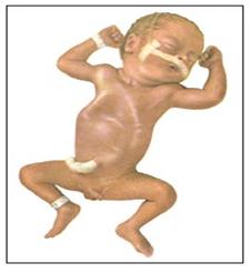
그림 308.특발성 호흡곤란 증후군이 아주 심하면 그로 인해 이 정도로 심한 호흡곤란이 생길 수 있다. 복벽과 흉곽 벽이 심하게 수축되어 흉곽 복벽 함몰 현상이 생겼다. 이런 호흡곤란 징후는 신생아 폐렴이 있을 때도 생길 수 있다.

Used with permission from Ross Laboratories, Columbus, Ohio 43216 Division of Laboratories, USA 과 소아가정간호백과

특발성 호흡곤란 증후군(특발성 호흡장애 증후군/유리질막증)의 증상 징후
  • 이 병의 정도, 합병증, 미숙 신생아의 체중 등에 따라 증상 징후가 다르다.
  • 이 병에 걸리면 미숙 신생아나 만삭 신생아에게 출생 후 곧 다음과 같은 여러 가지 증상 징후가 나타나는 것이 보통이다.
    • 1분에 60번 이상 숨을 자주 쉬는 것이 보통이고 때로는 1분에 60∼120회 정도 자주 쉬기도 한다.
    • 심한 경우 생후 곧 질식 상태에 빠질 수 있다.
    • 피부가 창백하고 파래질 수 있다.
    • 전신 청색증이 나타날 수 있다.
    • 숨 쉴 때마다 신음소리를 내기도 하고,
    • 특히 숨을 내쉴 때마다 그런팅(Grunting/그렁거림) 호흡을 하고
    • 주위 사람이 그런팅 호흡소리를 맨 귀로 들을 수 있고
    • 청진기로도 들을 수도 있다.
    • 비익이 숨 쉬는 대로 벌렁거리기도 한다.
    • 숨 쉴 때마다 가슴 늑골 사이(늑간)에 있는 늑골 근육이 흉강 속 쪽으로 빨려 들어갔다 나왔다 할 때도 있다.
    • 늑골 하 근육이 흉강 속 쪽으로 빨려 들어갔다 나왔다 할 수도 있고 늑골상위 근육도 흉강 속 쪽으로 빨려 들어갔다 나왔다 할 때도 있다(사진 308참조).
    • 젖이나 인공영양 젖병 꼭지를 제대로 빨지도 못한다.
    • 이 때 혈중 산소 농도는 정상 이하로 낮고(보통 PaO2가 55mm Hg이다),
    • 이산화탄소의 농도가 정상 이상으로 증가된다.
    • 이 병이 심할 때 조속히 적절하게 치료하지 않으면 생후 24∼48시간 이내에 호흡부전증 등으로 사망할 수 있다.
특발성 호흡곤란 증후군(특발성 호흡장애 증후군/유리질막증)의 진단 치료  
  • 병력·증상 징후 ·진찰소견 등을 종합해서 이 병이 의심되면 가슴 X선 사진을 찍어 진단한다. 
    • 선천성 후비공 폐쇄증, 
    • 기관 협착증, 
    • 기관지 협착증, 
    • 설 갑상선, 
    • 태변 흡인증, 
    • 일시적 빈호흡증, 
    • 박테리아 폐렴, 
    • 바이러스 폐렴, 
    • 기흉, 
    • 무기폐, 
    • 폐 발육부전, 
    • 선천성 심장 기형, 
    • 선천성 횡격막 허니아, 
    • 다 적혈구증, 
    • 기관 식도 누관(루), 
    • 두개강 내 출혈, 
    • 심낭 기종증, 
    • 저혈당증, 
    • 산혈증, 
    • 신생아 가사증, 
    • 쇼크 등으로 생긴 증상 징후와 특발성 호흡곤란 증후군으로 생긴 증상 징후가 비슷할 때도 있다. 그런 병들과 감별 진단해야 한다. 
  • 가슴 X선 사진, 초음파 검사, MRI 검사, Xenon 스캔 검사, 심장 에코그람 검사, 동맥혈 가스 검사, 혈청 포도당 검사, CBC 피 검사, 그 외 다른 적절한 임상 검사를 해서 진단한다. 
  • 10% 포도당 전해질용액 혈관주사로 수분과 영양분을 공급해 준다. 
  • 여러 가지 방법으로 산소호흡 치료를 하고 체온을 보온 해준다. 
  • 때로는 기관 내 내관 삽입치료를 한다. 
  • 기관 속에 기관 내 내관을 넣고 그 관을 통해 산소호흡 치료를 하기도 한다. 
  • 산소호흡 치료를 할 때 펄스 옥시메트리(Pulse oximetry) 검사나 동맥혈 피검사로 혈중 산소 농도를 수시로 측정한다. 
  • 요즘 펄스 옥시메트리 산소 농도측 정기로 동맥혈을 뽑지 않고 혈중 산소 농도를 손가락 등에서 쉽게 측정할 수 있다. 
  • 임신 32주 이후 출생한 비교적 큰 미숙 신생아들에게 생긴 특발성 호흡곤란 증후군은 적절히 치료하면 거의 다 회복될 수 있다. 
  • 이 병을 앓는 중 저산소증, 저혈당증, 빈혈, 기흉, 무기폐 등의 합병증이 생길 수 있다. 
  • 이 병은 항생제로 치료되지 않으나 서팩탄트제(표면활성제) 등으로 치료할 수 있다. 

Idiopathic respiratory distress syndrome in newborn infants (IRDS)

See the following information – www.drleepediatrics.com – Volume 6 Growth and Development and Nurturing Disorders Chapter 26: Respiratory Disorders in Newborns Please visit www.drleepediatrics.com-Volume 6 growth development parenting diseases -chapter 26: newborn respiratory disease
  201.jpg
Photo 307.
A newborn baby born prematurely has difficulty breathing and is receiving oxygen respiration treatment with an oxygen tube. After that, she was diagnosed with idiopathic respiratory distress syndrome and was treated. Copyright ⓒ 2011 John Sangwon Lee, M.D., FAAP
• The lungs of very small premature babies are more immature and have many physiological and functional features compared to the lungs of healthy newborns born at full term. See post-lens fibrosis.
• The alveoli of premature infants do not normally contain enough biochemical substances called Surfactant.
• Alveoli and bronchioles (細气管支/capillary bronchi) are also immature and their functions are immature.
• Oxygen in the inhaled air and carbon dioxide in the body are not properly exchanged in the alveoli of premature infants.
• For these reasons, the lungs of very small premature newborns are particularly prone to various diseases, including idiopathic respiratory distress syndrome.
• Among diseases caused by the immaturity of the lungs, vitreous membrane disease (idiopathic respiratory distress syndrome) is more likely to occur in premature newborns.
• In the past, idiopathic respiratory distress syndrome was the leading cause of death among premature infants.
• Studies have shown that 60% of premature newborns born before 28 weeks of gestation, 15-20% of premature newborns born between 32 and 36 weeks of gestation, and 5% of newborns born after 37 weeks of gestation have developed the disease.
• However, healthy full-term newborns born after 38 weeks of gestation rarely develop the disease.
• Today, the number of newborns with this disease is significantly lower than before.
• This disease is usually cured with good treatment.
• Premature newborns born to pregnant women with type 1 diabetes or preterm infants born by cesarean section are more likely to develop the disease.
  202.jpg
Figure 308. Idiopathic Respiratory Distress Syndrome, if very severe, can cause breathing difficulties of this magnitude.
Abdominal wall and thoracic wall were severely contracted, resulting in thoracic-abdominal wall depression.
These signs of shortness of breath may also occur with neonatal pneumonia. Used with permission from Ross Laboratories, Columbus, Ohio 43216 Division of Laboratories, USA Department of Pediatric and Family Nursing Encyclopedia
Symptoms of Idiopathic Respiratory Distress Syndrome
• Symptoms vary depending on the severity of the disease, complications, and the weight of the premature newborn.
• With this disease, it is common for premature or full-term newborns to show several symptoms and signs soon after birth, including:
o It is common to breathe more than 60 times per minute, and sometimes about 60 to 120 breaths per minute.
o In severe cases, you may fall into a state of suffocation soon after birth.
o The skin may be pale and blue.
o Systemic cyanosis may occur.
o Moaning with every breath,
o Grunting, especially with each exhalation
o People around you can hear grunting breathing with their bear ears
o Can also be heard with a stethoscope.
o The wing flaps as it breathes.
o Every time you breathe, the rib muscles located between the ribs of the chest (intercostals) are sucked in and out of the chest cavity.
o The subcostal muscles may be sucked in and out of the chest cavity, and the upper costal muscles may also be sucked in and out of the chest cavity (see photo 308).
o Inability to properly suck on nipples from milk or artificial nutrition bottles.
o At this time, the blood oxygen concentration is below normal (usually PaO2 is 55mm Hg), o The concentration of carbon dioxide increases above normal.
o If this disease is not treated promptly and properly when the disease is severe, it may result in death from respiratory failure within 24 to 48 hours after birth. Diagnostic treatment of idiopathic respiratory distress syndrome (idiopathic respiratory syndrome/vitreous membrane disease)
• If the disease is suspected based on medical history, symptoms, signs, and examination findings, a chest X-ray is taken for diagnosis.
o Congenital posterior nasal atresia;
o Tracheal stenosis;
o bronchial stenosis;
o lingual thyroid, o meconium aspirate;
o Transient tachypnea;
o bacterial pneumonia;
o viral pneumonia; o Pneumothorax, o atelectasis;
o Lung aplasia;
o Congenital heart malformations;
o Congenital diaphragmatic hernia; o polycythemia,
o Tracheal esophageal fistula (fistula); o intracranial hemorrhage,
o Pericardial emphysema; o hypoglycemia;
o Acidemia; o Neonatal dyslexia;
o Symptoms and signs caused by shock and the like are sometimes similar to those caused by idiopathic respiratory distress syndrome. It should be diagnosed differently from such diseases.
• Diagnosis is made by performing chest X-ray, ultrasound, MRI, Xenon scan, cardiac echogram, arterial blood gas test, serum glucose test, CBC blood test, and other appropriate clinical tests.
• Water and nutrients are supplied by vascular injection of 10% glucose electrolyte solution.
• Oxygen breathing therapy is performed in various ways and the body temperature is kept warm.
• Sometimes endotracheal tube insertion is performed.
• An endotracheal tube is inserted into the trachea and oxygen respiration is sometimes performed through the tube.
• During oxygen respiration therapy, the blood oxygen concentration is frequently measured with a pulse oximetry test or an arterial blood test.
• Nowadays, with a pulse oximetry oxygen concentration meter, it is possible to easily measure the blood oxygen concentration on a finger, etc. without drawing arterial blood.
• Idiopathic respiratory distress syndrome in relatively large premature newborns born after 32 weeks of gestation is almost completely reversible with proper treatment.
• During this disease, complications such as hypoxia, hypoglycemia, anemia, pneumothorax, and atelectasis may occur.
• This disease cannot be treated with antibiotics but can be treated with surfactants (surfactants).


출처 및 참조 문헌 Sources and references

  • NelsonTextbook of Pediatrics 22ND Ed
  • The Harriet Lane Handbook 22ND Ed
  • Growth and development of the children
  • Red Book 32nd Ed 2021-2024
  • Neonatal Resuscitation, American Academy Pediatrics
  • www.drleepediatrics.com 제1권 소아청소년 응급 의료
  • www.drleepediatrics.com 제2권 소아청소년 예방
  • www.drleepediatrics.com 제3권 소아청소년 성장 발육 육아
  • www.drleepediatrics.com 제4권 모유,모유수유, 이유
  • www.drleepediatrics.com 제5권 인공영양, 우유, 이유식, 비타민, 미네랄, 단백질, 탄수화물, 지방
  • www.drleepediatrics.com 제6권 신생아 성장 발육 육아 질병
  • www.drleepediatrics.com제7권 소아청소년 감염병
  • www.drleepediatrics.com제8권 소아청소년 호흡기 질환
  • www.drleepediatrics.com제9권 소아청소년 소화기 질환
  • www.drleepediatrics.com제10권. 소아청소년 신장 비뇨 생식기 질환
  • www.drleepediatrics.com제11권. 소아청소년 심장 혈관계 질환
  •  www.drleepediatrics.com제12권. 소아청소년 신경 정신 질환, 행동 수면 문제
  • www.drleepediatrics.com제13권. 소아청소년 혈액, 림프, 종양 질환
  • www.drleepediatrics.com제14권. 소아청소년 내분비, 유전, 염색체, 대사, 희귀병
  • www.drleepediatrics.com제15권. 소아청소년 알레르기, 자가 면역질환
  • www.drleepediatrics.com제16권. 소아청소년 정형외과 질환
  • www.drleepediatrics.com제17권. 소아청소년 피부 질환
  • www.drleepediatrics.com제18권. 소아청소년 이비인후(귀 코 인두 후두) 질환
  • www.drleepediatrics.com제19권. 소아청소년  안과 (눈)질환
  • www.drleepediatrics.com 제20권 소아청소년 이 (치아)질환
  • www.drleepediatrics.com 제21권 소아청소년 가정 학교 간호
  • www.drleepediatrics.com 제22권 아들 딸 이렇게 사랑해 키우세요
  • www.drleepediatrics.com 제23권 사춘기 아이들의 성장 발육 질병
  • www.drleepediatrics.com 제24권 소아청소년 성교육
  • www.drleepediatrics.com 제25권 임신, 분만, 출산, 신생아 돌보기
  • Red book 29th-31st edition 2021
  • Nelson Text Book of Pediatrics 19th- 21st Edition
  • The Johns Hopkins Hospital, The Harriet Lane Handbook, 22nd edition
  • 응급환자관리 정담미디어
  • Pediatric Nutritional Handbook American Academy of Pediatrics
  • 소아가정간호백과부모도 반의사가 되어야 한다이상원 저
  • The pregnancy Bible. By Joan stone, MD. Keith Eddleman, MD
  • Neonatology Jeffrey J. Pomerance, C. Joan Richardson
  • Preparation for Birth. Beverly Savage and Dianna Smith
  • 임신에서 신생아 돌보기까지이상원
  • Breastfeeding. by Ruth Lawrence and Robert Lawrence
  • Sources and references on Growth, Development, Cares, and Diseases of Newborn Infants
  • Emergency Medical Service for Children, By Ross Lab. May 1989. p.10
  • Emergency care, Harvey Grant and Robert Murray
  • Emergency Care Transportation of Sick and Injured American Academy of Orthopaedic Surgeons
  • Emergency Pediatrics A Guide to Ambulatory Care, Roger M. Barkin, Peter Rosen
  • Quick Reference To Pediatric Emergencies, Delmer J. Pascoe, M.D., Moses Grossman, M.D. with 26 contributors
  • Neonatal resuscitation Ameican academy of pediatrics
  • Pediatric Nutritional Handbook American Academy of Pediatrics
  • Pediatric Resuscitation Pediatric Clinics of North America, Stephen M. Schexnayder, M.D.
  • Pediatric Critical Care, Pediatric Clinics of North America, James P. Orlowski, M.D.
  • Preparation for Birth. Beverly Savage and Dianna Smith
  • Infectious disease of children, Saul Krugman, Samuel L Katz, Ann A.
  • 4권 모유모유수유이유 참조문헌 및 출처
  • 5권 인공영양우유, 이유, 비타민, 단백질지방 탄수 화물 참조문헌 및 출처
  • 6권 신생아 성장발육 양호 질병 참조문헌 및 출처
  • 소아과학 대한교과서

Copyright ⓒ 2014 John Sangwon Lee, MD., FAAP

부모도 반의사가 되어야 한다”-내용은 여러분들의 의사로부터 얻은 정보와 진료를 대신할 수 없습니다.

“The information contained in this publication should not be used as a substitute for the medical care and advice of your doctor. There may be variations in treatment that your doctor may recommend based on individual facts and circumstances.

“Parental education is the best medicine.